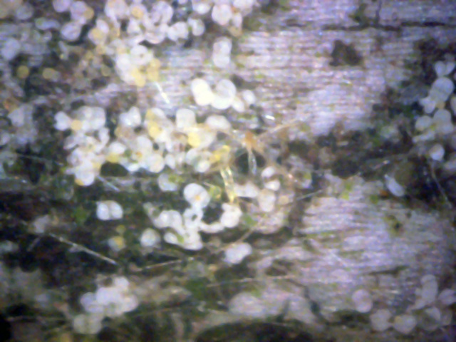
I spotted a large sick looking Honey Locust tree about a mile from my house.
Its foliage was somewhat thinner than normal and many of the lower branches were dead.
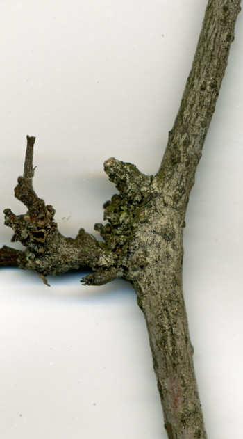
In early June I removed the tip of one of these dead branches.
It was about a quarter inch thick and is shown in the first picture below.
1
The tan discoloraton almost appears as if it were spray painted on. In fact,
this discoloration is due to a large number of tiny spores. They were found
almost exclusively on one side of the branch.
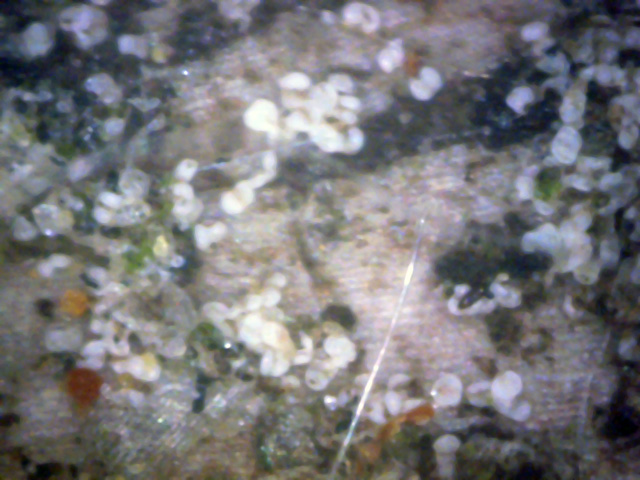
3
The above two pictures are 400x digital microscope views of the tan area at the
branch junction shown in picture 1. The white objects look exactly like the
white canker spores seen on other trees and shrubs.
In early October, hoping to gather more conclusive evidence of white canker infection,
I returned to this tree to obtain a branch that I could examine with a digital microscope.
The following set of pictures confirm the white canker infection. The first
three pictures are of leaf tops.
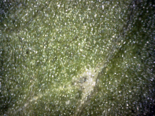
4
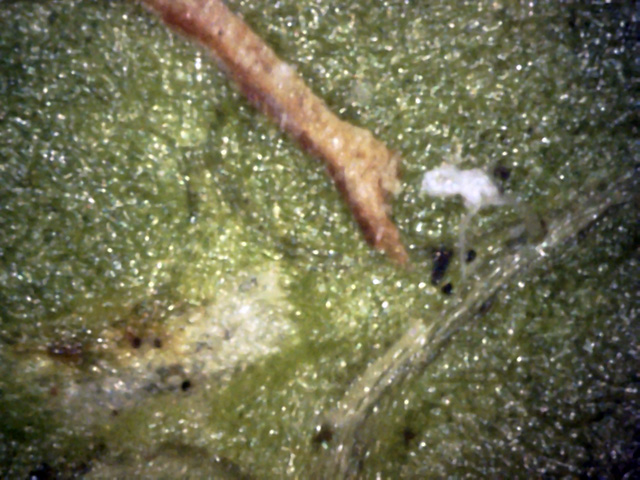
There wasn't much evidence of infection on the leaf's top, as shown here.
Only occasional white areas like this gave a hint of the canker within.
5
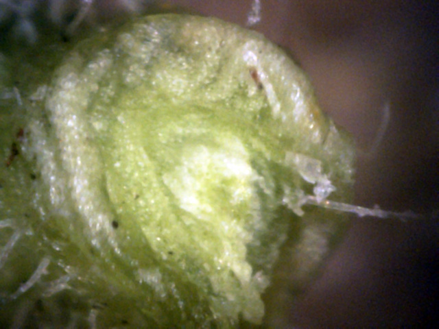
These two areas show a bit more evidence of white canker infection.
The canker on the left is probably below the surface, but the one on the right
is on the surface - you can just make out the stalk where it connects to a leaf vein.
The brown stick-like object is probably another infection.
 ←
↑
←
↑
6
This isn't a leaf top, but the torn end of the leaf stem.
The crystal clear tissue on the right (red arrows) probably indicates that white canker
destroyed all the chlorophyll.
Usually leaf bottoms are an even better indicator of white canker infections that leaf tops.
That's not quite true here, as shown in these microscope leaf bottom pictures.
 ↑
↑
7
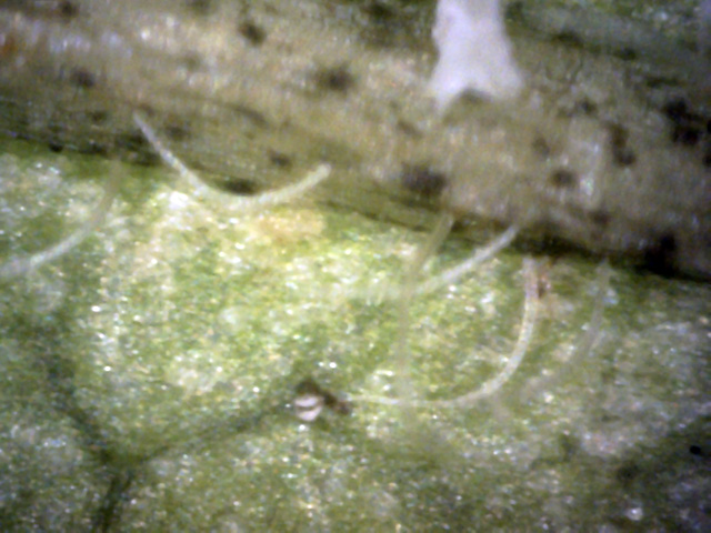
Surprisingly, the leaf bottom was fairly clean.
Only near the stem were there any hints of infection.
Here there is a blob of canker material on the stem.
There is also what appears to be a dead white canker spore (red arrow).
 ←
←
←
←
8
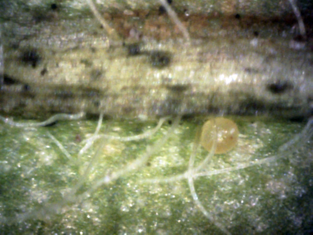
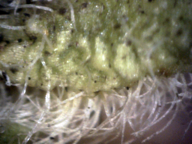
The leaf stem with lots of hyphae growing from it. But note the yellow sphere.
It appears to be connected to the stem with a barely discernable hypha (red
arrow),
so it probably isn't an insect egg.
In addition, if you look closely, the stem hypha in the upper left (blue arrow) appears to be
developing a yellow swelling at its base.
9
As we get close to stem at the base of the leaf, the number of hyphae proliferate like grass.
But the white blobs under the leaf surface make one wonder if the blobs aren't
canker responsible for this hyphae growth.
 ↑
↑
←
↑
↑
←
10
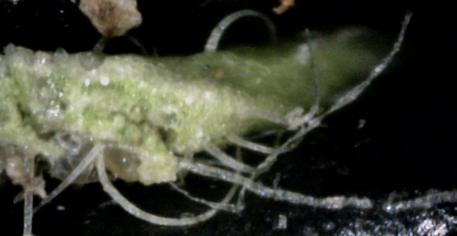
There are a lot of hyphae on a leaf vein, particularly near the base.
So here I did a cross-section of a leaf base where there were lots of hyphae.
This shows there is a difference between a normal leaf hair (red arrow) and a white canker hyphae
(blue arrow).
Both are semi-transparent. Leaf hair has a round cross-section, a smooth surface,
and gradually tapers to a point.
Canker hypha, in contrast, is more like a ribbon.
It has a 1:4 cross-section aspect ratio, twists, is lumpy, and tends to run for a
relatively long distance.
They also tend to have an occasional black spot and occasional tiny protrusions
(yellow arrow - small root-like structures).
Leaf cross-section views with a microscope will often give a good indication as
to whether white canker exists, and how pervasive it is. However, in the case of
honey locust trees, these views aren't that useful, as the following three
pictures show.
11
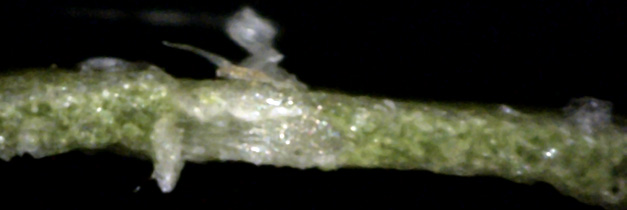
The leaf cross-sections actually show little of interest.
Here is an exception where canker extends both below and above the leaf surface.
12
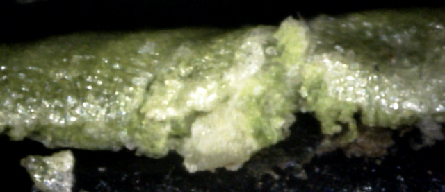
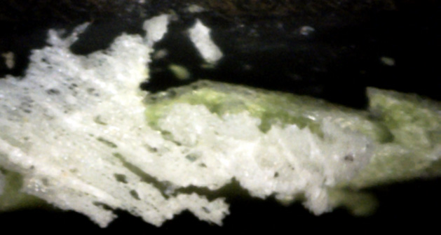
In the middle of this leaf cross-section, you can see a blob of canker.
However, its shape appears as if it might be a leaf vein that was consumed
by white canker.
13
Here the leaf's cross-section is hidden.
White canker will sometimes cover the leaf's surface and infect and kill
the surface cells.
Then, when a razor tries to slice through this tougher material, it instead lifts
it up as a sheet and drops it elsewhere, as was done here.
This action seems similar to a bad sunburn, where the surface skin cells die
and then later peel off in small sheets.
The most reliable white canker diagnostic views are twig cross-section views.
They have an added advantage in that they don't degrade over the course of a few days
or weeks, as leaves do. This reliable disgnostic evidence is confirmed by the 6 twig
cross-section pictures below, taken with a 400x digital microscope.
14
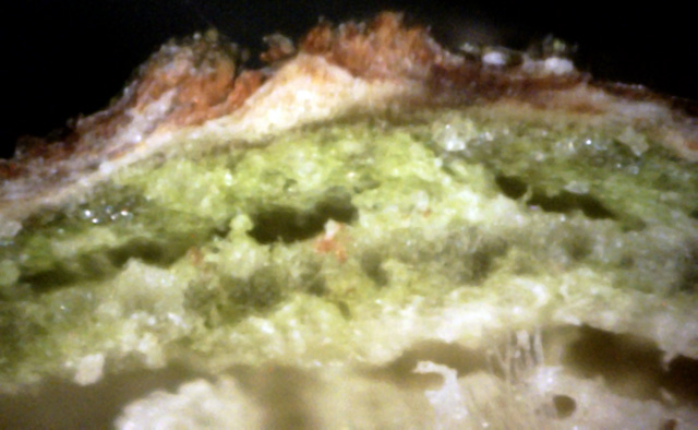
Extensive canker damage.
The phloem has a void in the middle of it and the dark green cortex is virtually gone.
Even the outer xylem has a large void!
Most telling of all, the top bark has such a significant canker growth
that it is bursting out of the bark and looks like a volcano.
15
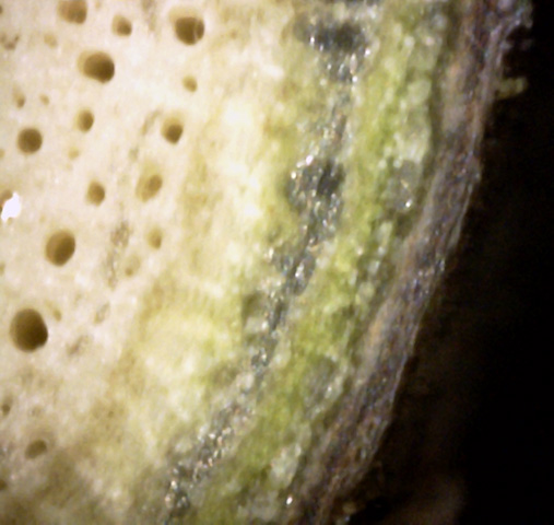
Reasonably healthy area area with intact outer bark.
But there is beginning to be a large amount of canker growth between the dark green
cortex and the phloem - lots of tan canker particles are building up there.
 Pith
Xylem
16
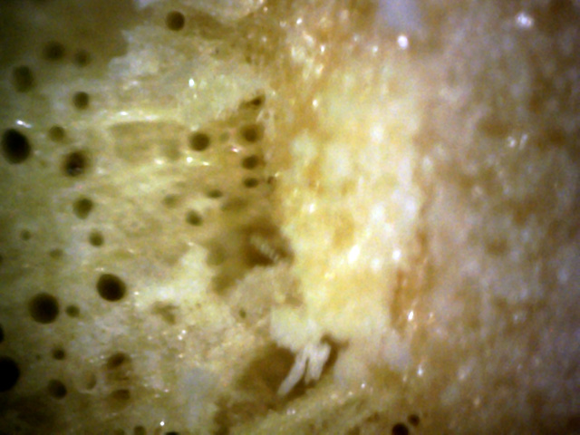
This is the junction of the pith (twig center)
and the xylem (area with all the vessels).
I usually ignore this area since there's not much of interest there,
but in this case the canker has grown here too.
 ←
←
←
←
←
←
←
←
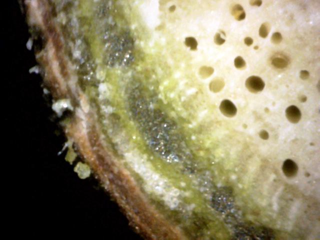
17
There is an extensive area of snow-white canker tubule growth here just under the bark.
But notice also the several areas where the canker is growing on the bark - it is
eating away the bark, blackening and destroying the surrounding area as it does so (blue arrows).
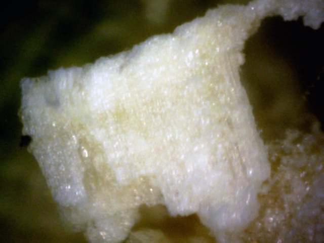
18
White canker appears to be tougher than regular wood, so when a cut is made through
the wood, it will sometimes dislodge a solid chunk of this canker.
Apparently that is what happened here, giving us a good view of
pure white canker.
 ↓
←
←
↓
↓
←
←
↓
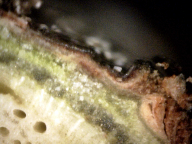
19
Lots of information in this view.
On the far right is a major battle between the white canker and the tree's bark,
causing an eruption through the bark, with clear white canker bubbling out the top
(red arrow).
In the middle of this picture is an infected area of white canker, indicated by the
white particles (blue arrow).
Interestingly, underneath it we can see that it has also infected the rays, which
look like white nails (green arrow). And above it, the canker appears to be pushing the bark out a bit.
White canker has even broken through on the outer bark, as seen by the out-of-focus
white particles on the bark's surface (yellow arrow).
